

Breakthrough Near-Infrared Holography Pushes Lensless Microscopy Beyond Its Traditional Visible-Range Scattering Limit
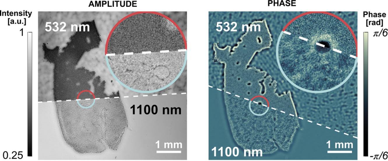
Near-Infrared Holography and Extended Sample-Sensor Distance Enhance Lensless Imaging

QCI Lab team
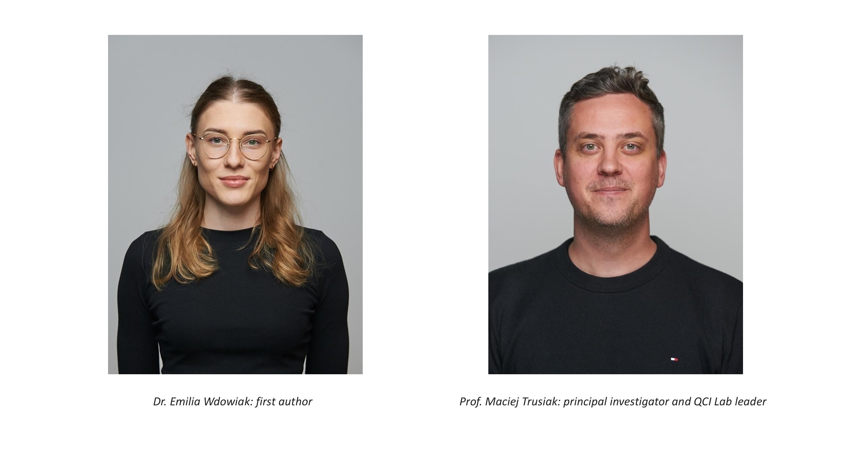
Dr. Emilia Wdowiak: first author | Prof. Maciej Trusiak: principal investigator and QCI Lab leader
Near-infrared light and a larger sample-sensor gap recover detail where classical in-line visible-range lensless holography fails due to strong scattering
CHENGDU, SICHUAN PROVINCE, CHINA, October 16, 2026 /EINPresswire.com/ -- Near-infrared light and an unexpectedly larger sample-sensor gap recover detail where classical in-line visible-range lensless holography fails due to strong scattering - Report about Articles Published in Opto-Electronic AdvancesLensless holographic microscopy replaces imaging optics with computation, but its classical Gabor principle has long been confined to thin, weakly scattering specimens. Researchers at Warsaw University of Technology have now pushed the method into the multiple-scattering regime. Near-infrared illumination preserved resolvable information through tissue-like scattering layers up to 1.4 mm, while increasing the sample-sensor distance unexpectedly doubled lateral resolution—revealing a new acquisition strategy for high-throughput, robust, amplitude-and-phase imaging in complex media.
Light scattering is one of microscopy's oldest barriers. In a transparent specimen, light follows paths that can be traced back to structure. In thick, heterogeneous tissue, however, photons are redirected again and again, scrambling the information. The image-forming signal loses power, fine detail disappears, and even sophisticated reconstruction can be left with little more than haze. Conventional microscopes can recover depth by adding specialized optics, scanning, labels, or tissue clearing, but each intervention costs time, field of view, or instrument complexity.
Lensless digital holographic microscopy approaches the problem from a radically different direction. The specimen is placed close to a bare image sensor, with no objective lens between them. A compact source illuminates the sample, and the sensor records an interference pattern rather than a conventional photograph. Numerical propagation then reconstructs locally both amplitude—how strongly the specimen attenuates the light—and phase—how much it delays the wave. Because no microscope objective narrows the scene, the entire sensor becomes the field of view: about 116 mm2 in this study, captured without raster scanning or image stitching.
This simplicity has always concealed a fundamental weakness. Gabor's in-line holography assumes that a strong, unscattered reference wave interferes with a much weaker object wave. The assumption works well for isolated cells and very thin, transparent specimens. Once multiple scattering becomes strong, the reference is depleted, holographic fringes are buried in diffuse background, and reliable reconstruction breaks down. The method has therefore remained largely on the weak-scattering side of microscopy.
The Warsaw University of Technology team and cooperators, led by Professor Maciej Trusiak, asked whether this boundary was as fixed as it appeared. Near-infrared light is redirected less strongly by many tissue structures than visible light, allowing more useful information to survive propagation through a turbid sample. In a conventional microscope, exploiting that advantage can require infrared detectors, lenses, and coatings. In a lensless system, there are no imaging lenses to replace—and even the faint residual response of an ordinary silicon sensor near 1100 nm may be enough for holographic reconstruction. The idea was disarmingly simple: change the wavelength, preserve the architecture, and test whether the lost image information could be made visible again via invisible near-infrared wave.
The research group of Prof. Trusiak at the Quantitative Computational Imaging Lab (QCI Lab), Institute of Micromechanics and Photonics, Warsaw University of Technology, Poland, tested this idea across six illumination wavelengths from 480 to 1100 nm without changing the core imaging platform. Tissue-mimicking milk layers provided controlled and repeatable levels of scattering, while calibrated targets measured the finest features that remained recoverable. The findings of the study were published online in the journal Opto-Electronic Advances on September 20, 2026.
The result was a decisive spectral shift in performance. Visible-light operation became fully unresolved at scattering-layer thicknesses between 180 and 360 µm, depending on wavelength. At 1100 nm, resolvable features survived through approximately 1.4 mm of the tissue-like medium—nearly four times the thickness reached at 632 nm. Remarkably, the standard visible-range CMOS sensor achieved robust reconstructions even though its quantum efficiency at 1100 nm was just 0.19%. The experiment shows that residual sensitivity, usually treated as negligible, can become useful when paired with a low-photon-budget holographic method.
“The study also overturned one of the field's practical instincts. Lensless systems normally place the sample as close to the sensor as possible to maximize the resolution. Under strong scattering, the team found that increasing this distance from about 3 to 12 mm improved lateral resolution from roughly 11 to 5.5 µm,” says Prof. Trusiak. The proposed explanation is that useful interference-carrying light remains more concentrated than the broadly diffused background; with additional propagation, the noise spreads faster, making fringes easier to detect. The effect still requires deeper mechanistic study, but it immediately reframes sample-sensor distance as a parameter to optimize rather than simply minimize.
Most importantly, the controlled experiments translated to biology. At 1100 nm, the system produced ultra-wide-field, label-free amplitude, and phase images of uncleared mouse liver sections up to 60 µm and brain sections up to 250 µm, revealing internal organization that visible illumination largely obscured. Rather than claiming non-invasive imaging deep inside a living organ, the advance establishes a new operating regime for compact in vitro holography: rapid, wide-area recovery of intrinsic tissue structure in thick, strongly scattering sections. “Future work can combine distance-aware acquisition with scattering-aware or physics-informed reconstruction and more sensitive detectors, pointing toward faster tissue survey, computational imaging through complex media, and compact instruments for laboratories where conventional platforms are difficult to scale,” says Prof. Trusiak.
QCI Lab, led by Prof. Trusiak develops optical methods in which computation becomes part of the microscope. Its work spans quantitative phase imaging, digital holography, lensless microscopy, and holographic tomography, with a consistent goal: extract rigorous biological information via advanced computations from compact, scalable hardware. The group has contributed lensless methods for e.g., suppressing holographic twin images, reconstructing data recorded with very limited photon budgets, and creating gigavoxel-scale three-dimensional maps of biological specimens.
The present study was led by first author Dr. Emilia Wdowiak from the Warsaw University of Technology, Poland, and brought optical engineering together with biological and clinical expertise from the Laboratory of Neurobiology at the Nencki Institute of Experimental Biology, Polish Academy of Sciences, Poland; the Department of Immunology at the Medical University of Warsaw, Poland; the Department of Pathology at the Maria Sklodowska-Curie National Research Institute of Oncology, Poland; and the Centre of Postgraduate Medical Education, Poland. “This collaboration enabled the team to move beyond resolution targets and test the method on biologically meaningful tissue architecture,” says Dr. Wdowiak.
Reference
Title of original paper: Near-infrared lensless holographic microscopy on a standard sensor for label-free mesoscale imaging in multiple scattering
Journal: Opto-Electronic Advances
DOI: https://doi.org/10.29026/oea.2026.260088
Funding information
The research was supported by Poland's National Centre for Research and Development through the LIDER XIV programme. Equipment was co-funded by the Warsaw University of Technology Excellence Initiative: Research University (IDUB) programme.
Siyi Ma
Institute of Optics and Electronics
+86 191 3618 6296
Legal Disclaimer:
EIN Presswire provides this news content "as is" without warranty of any kind. We do not accept any responsibility or liability for the accuracy, content, images, videos, licenses, completeness, legality, or reliability of the information contained in this article. If you have any complaints or copyright issues related to this article, kindly contact the author above.
